Microbiology Lab
Exercise 10: Smear
Preparation
Exercise 11:
Simple Staining
Ex 12: Negative
Staining
REVIEW LAB RESULTS FROM LAST WEEK
- Ex 8: Aseptic Technique
- Did you
successfully get bacteria growing in your broth and slant?
- Ex 6: The Ubiquity of Bacteria
- Which plate did
you expect to have the worst contamination?
- Which plate
actually had the worst contamination?
- What surprised
you the most about the experiment?
Ex 10:
Smear Preparation
- Every student
will prepare 2 slides of E. coli
(1 from the slant & 1 from the broth started by students last week)
for use in the simple staining lab.
- Procedure
- Gather supplies
i. Bacterial cultures (1
slant & 1 broth of E. coli)
ii. Clean microscope
slides
iii. Inoculating loop
iv. Water bottle
v. Sharpie pen
vi. Bunsen burner &
striker
- Label slide
i. Label a short edge of
the slide with the bacteria, your initials, & date
ii. Draw a target circle
about ½” wide
iii. Turn your slide over
so you work on the unlabeled side
- Aseptically
collect a sample of your bacteria
i. When you are using
broth, transfer two loopfuls of bacteria to your slide
ii. When you are suing
solid media, place 2 loopfuls of water on the slide, then transfer a SMALL
amount of organisms to your slide
- Spread your
organisms out over the entire area of your target circle
- Let the smear
air dry
- Heat fix the
slide by passing it through the flame of a Bunsen burner until the slide
is very warm to the touch
Ex 11:
Simple Staining
·
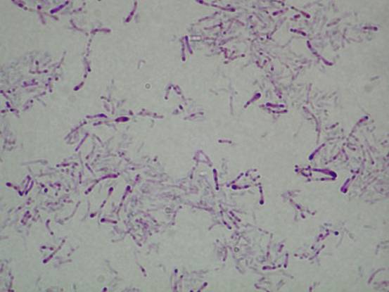
Look
at the view of Corynebacterium
diphtheriae at
www.mf.uni-lj.si/imi/images/preparati/mikrobi/coryn01.jpg
o
I
went to google & searched “Corynebacterium
diphtheriae images”
o
This
slide was prepared by…
§ Preparing a smear.
§ Staining with
methylene blue for one minute
o
Note
the following in this view…
§ Pleomorphism – irregularity of form
·
C. diphtheriae is a bacillus, but
may look like a rod, club-shaped, spermlike, or needle-shaped
§ Metachromatic granules
§ Palisade arrangement – a “picket fence” arrangement
·
Prepare
a simple stain of Escherichia coli
o
Use
your prepared smear of E. coli
o
Stain
with methylene blue for one minute
o
Rinse
with water
o
Blot
with bibulous paper
o
Observe
under oil immersion lens
·
What
is the morphology and arrangement of E.
coli?
|
Gram Stain of E. coli |
|
|

Ex 12:
Negative Staining of Staphylococcu
aureus and Bacillus megaterium
·
Negative stains – stains that don’t
penetrate the bacterial cell wall, but cause the background slide to appear
dark while the bacteria remain colorless
·
Prepare
2 negative stain slides: 1 each of S. aureus and B. megaterium
o
Label
the slide with the bacteria’s name, your initials, & date
o
Place
a loopful of India ink in the center of your slide
o
Aseptically
collect a loopful of bacteria and transfer them to the slide with the India ink
o
spread
the organisms out over an area of about ¾”
o
Allow
to air dry
o
View
under oil immersion
·
What
is the morphology & arrangement of S.
aureus?
·
What
is the morphology & arrangement of B.
megaterium?
|
Negative stain of
B. megaterium (note the
endospores that don’t take up the
stain) |
Gram stain of S. aureus |
|
|
|

